
Laurence is a copywriter and trained journalist. His passion is creating insightful and educational content for dental professionals of every level. Outside of work, he enjoys watching sport, quizzing and spending time with his family.
You May Also Like
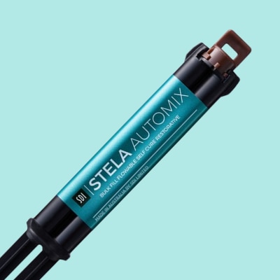
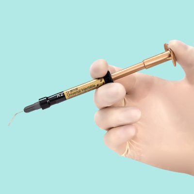
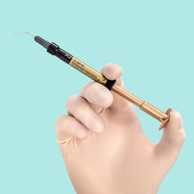
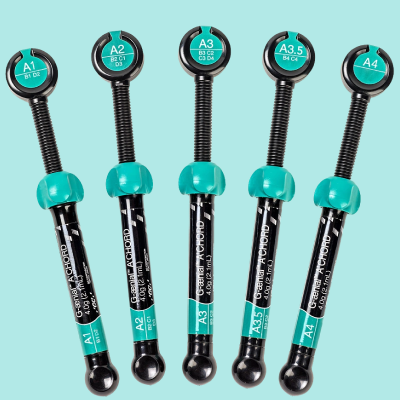
SDI Stela
Stela is the ideal option for dentists seeking a strong and reliable high-performance restorative, that is simple, fast and does not compromise aesthetics.
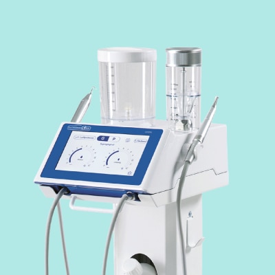
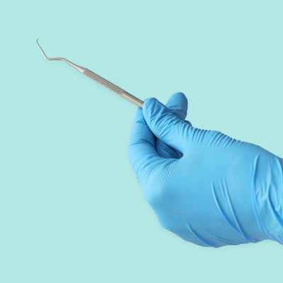
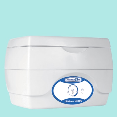
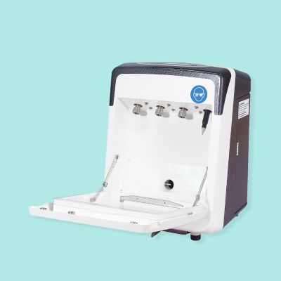
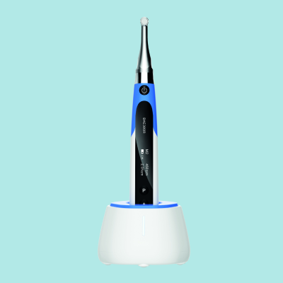
The Ultimate In Scale and Polish
The UC500L has been designed to make scale and polish treatments easier and more efficient for you and more comfortable for your patients.
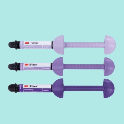
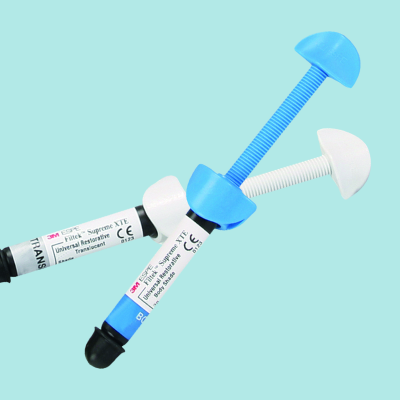
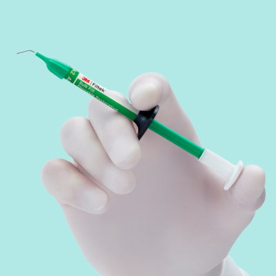
Filtek Easy Match Restorative
Filtek Easy Match Universal Restorative is a 3-shade system designed to deliver an excellent shade to almost any patient’s tooth shade.
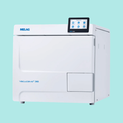
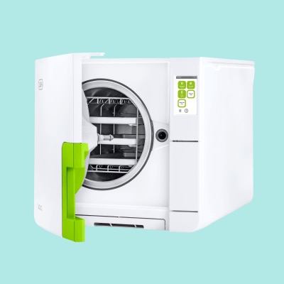
Melag Autoclaves
Melag autoclaves and steam sterilizers for practices and clinics. Providing sterilisation in compliance with international standards.
Amalgam Alternatives from GC
Whether your preference is for composite or GIC, GC is a brand that specialises in easy-to-use restoratives that can help smooth your amalgam phase-down.
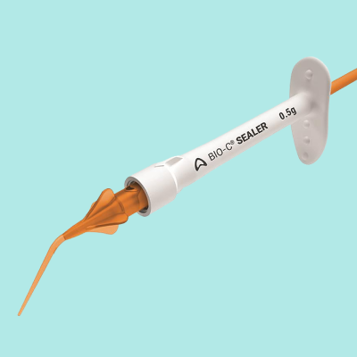
Angelus Root Canal
All products in the Angelus root canal sealers and cements range are designed to improve the workflow of DCPs and patient outcomes.

SDI Exclusive Offers
Don't miss this limited-time opportunity to stock up on your favourite SDI products!
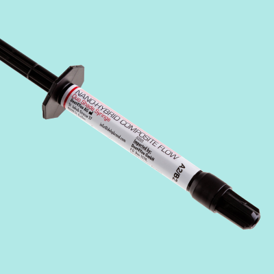
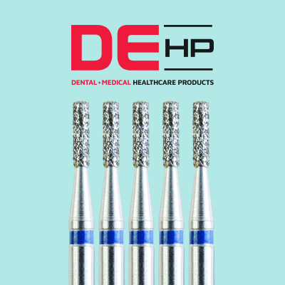
DEHP Nano Flowable
Find out how DEHP Nano Flowable Composite has stayed Kent Express' best-selling composite for several years running.
Enhance Your Restorations
Discover how 3M's impression materials, composites and polishing discs can optimise your restorations from start to finish.
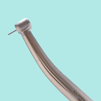
BA International Small Equipment
From curing lights and air polishers to autoclaves and handpiece lubrication units, BA International is a world leader in dental small equipment
Elements Connect & Apex Connect
When seamlessly connected with the Kerr Apex Connect apex locator, the elements Connect can bring added confidence and accuracy to all shaping procedures.
Importance of Decontamination
Accurate record keeping is an essential part of running a dental practice and is necessary in the provision of high-quality, safe and effective patient care.
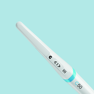
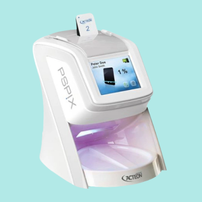
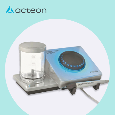
5 Reasons To Buy Acteon C50
With the continuous innovation in dental cameras for precise imaging and a greater emphasis on preventive care, intraoral cameras (IOCs) have never been more popular.

6 Reasons To Trust Kent Express
Discover just what makes Kent Express Dental Supplies the go-to e-commerce store for thousands of dentists every year.
Solutions for Root Caries
Root caries in older patients should be approached holistically with a treatment plan that identifies, protects, treats and controls.
Can Bubbles Be Avoided
How 3M Filtek Flowable Restorative can help remove the challenge of bubbles with its innovative syringe design other advantages.

Own Brand Essentials Checklist
Switching to Kent Express own brands for your everyday essentials can provide your practice with serious yearly savings.
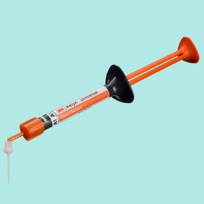
Universal Resin Cement Can Help
To simplify dental cementation try a universal resin cement, such as RelyX from 3M. Find out more.
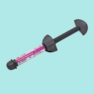
3M Restoratives
3M has several restorative materials designed to remove unnecessary stress at every stage of a restoration and make life easier.
Guide to Dental Scalers
Make your scaling procedures as efficient and effective as they can be with our extensive guide to dental scalers.
Enbio S Autoclave
The Enbio S autoclave from BA International is capable of sterilisation speeds 4 times faster than the average autoclave. Find out more.
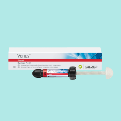
Kulzer Venus One Range
Your ONE shade solution for everyday cases. Enjoy efficient handling and long-lasting restoratives with Kuler Venus One composites.

Retraction Cord Vs Paste
A look at the pros and cons of the two most popular gingival retraction methods for impression taking prep

Enhance Composite Restorations
6 tips for success to help you get the most out of your flowable composite restoratives.
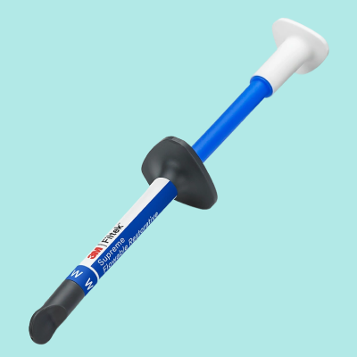
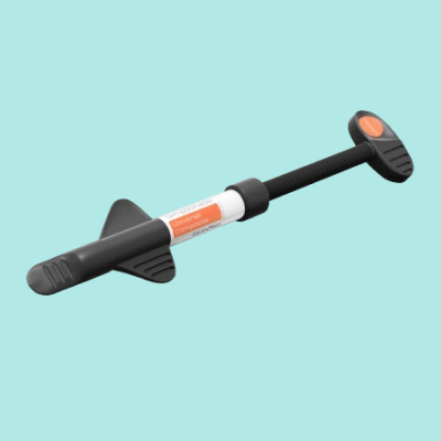
3M Filtek Supreme Flowable
3M Filtek Supreme Flowable Restorative and helps to virtually eliminate bubbles, cut waste and reduce the thumb pressure needed for injection.


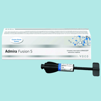
Admira Fusion 5
Admira Fusion 5 is the first purely ceramic-based restorative material without no classic monomers.


Best Value with BA Handpieces
3 great reasons why BA dental handpieces offer the best value for your dental practice.
Handpiece and Equipment Repairs
In partnership with B.A. International, Kent Express provide a fast handpiece and small equipment repair service.
Phosphor Plate Radiography
Phosphor plate radiography is considered by many the least expensive way to convert from analogue to digital imaging and have the easiest learning curve.

Avoid Voids in Impressions
Advice on the best techniques and materials to use to avoid those dreaded impression voids.
Consider Own Brand Restoratives
Try our 100% guaranteed Own Brand restoratives and discover products with near identical quality to big name brands but at a lower price.
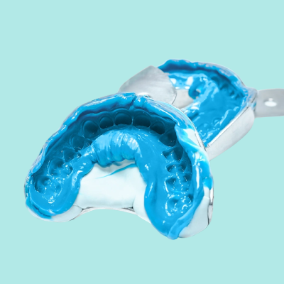
Silicone Impression Materials
An in-depth look at the similarities and key differences between the two main silicone impression materials
Guide to Ultrasonic Baths
Everything you need to know about the use of ultrasonic baths in dentistry and how to find the best ultrasonic cleaner for you.
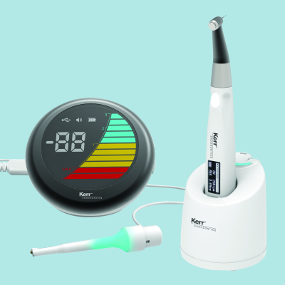
Endo Motors With Apex Locators
Having your apex locator built into your endo motor can streamline root canal procedures and reduce operation times.
Glass Ionomer Fillings
How switching to 3M glass ionomer products could make fillings and other procedures quicker, easier and less messy.

Why Do Dental Implants Fail
A look at why dental implants sometimes fail and the ways to minimise the chances of this happening.
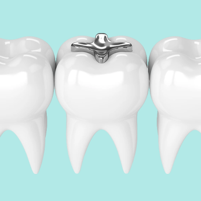
Amalgam Fillings in Dentistry
Could amalgam fillings be gone by 2030? We discuss the past, present and future of amalgam.
Periodontal Therapy
Taking a scientifically supported approach to periodontal treatment is key for successful outcomes.
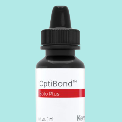
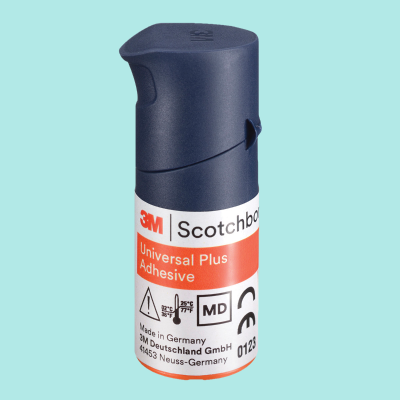
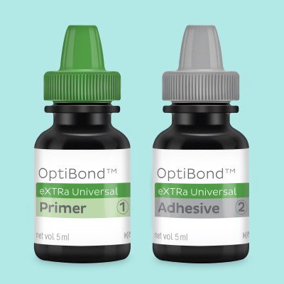
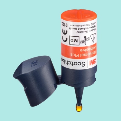
Kerr OptiBond Solo Plus
Kent Express’ best-selling total-etch adhesive for several years running, Kerr OptiBond Solo Plus offers qualities not found in other adhesive systems
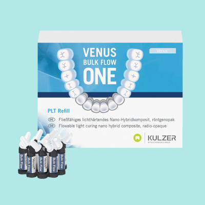
Benefits of Venus Bulk Flow ONE
With NHS dental appointments still unavailable to many, some have resorted to treating their teeth at home in what is being dubbed ‘DIY dentistry’.
Find the Best Prophy Paste
Finding a good prophy paste can make hygiene treatment a more comfortable and rewarding experience for dentists and patients alike.
Guide to Autoclaves
All the knowledge you need to make an informed decision on what autoclave to buy for your dental practice, including the key features to look out for.
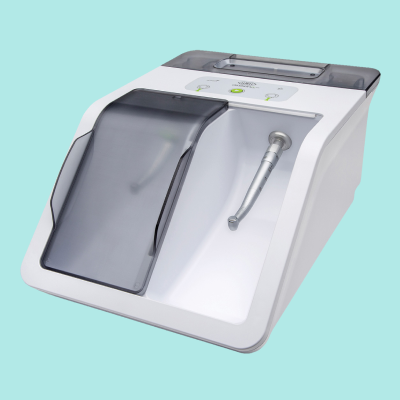
Why You Need a Lubrication Unit
The cutting-edge Assistina TWIN automatic handpiece maintenance device from W&H provides an exceptional, consistent standard of lubrication in just 10 seconds.
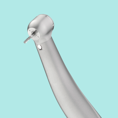
BA Ultimate Turbine
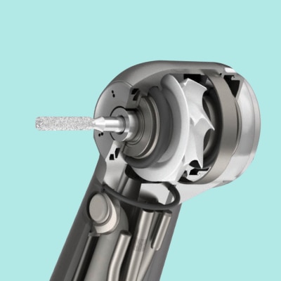
Looking for a reliable, low maintenance high speed turbine? The BA Ultimate range ticks both boxes and comes with several other outstanding features.
Relieving Stress, Composite Way
How 3M restoratives can help take stress out of composite bonding, placement, curing and shade matching.
Guide to Handpiece Maintenance
With some basic understanding of how dental handpieces work and how best to look after them, handpieces can maintain efficiency for longer.
Simplishade reduces chair time
How time management, not more patients, is the key to increased productivity and profitability in the dental practice.
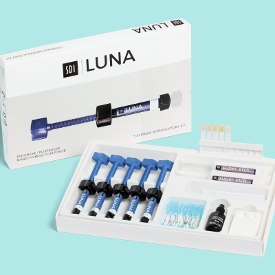
SDI Luna Nanohybrid Composite
Everything you need to know about the new SDI Luna nanohybrid composite and the first-hand experiences using it by a dental care professional.

Choosing GC Restorative Material
Fall in love with your next restorative material from GC. Discover the benefits of some of GC'S most popular composite and glass hybrid restoratives.
Restorative Treatments Guide
Our 4-Step guide to more efficient restorative treatments can help to make direct restorative treatment an easier and more effective process.
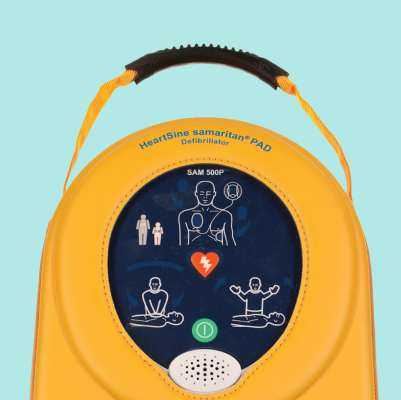
Importance of a Defibrillator
Learn why every dental practice should have a defibrillator with Kent Express informative guide.
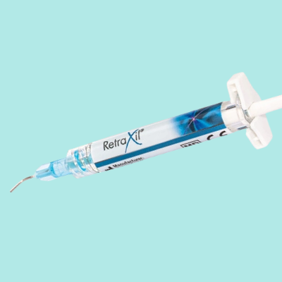
RetraXil Retraction Paste
Astringent retraction paste can provide several benefits over cords for patient and dentist. Advanced pastes such as RetraXil, are optimising these benefits
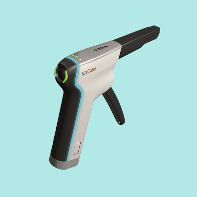
Voco VisCalor
Find out why Voco VisCalor one of the worlds most popular dental materials and how it is the world’s first universal composite to use thermoviscous technology
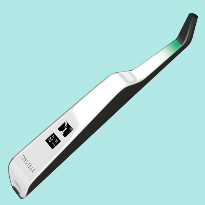
Simplify Implant Stability
We can help you simplify implant stability with the Osstell Beacon, it uses RFA to determine stability in seconds to help simplify and improve implant results.

Equipment to Lower Your Bills
Here is a look at some equipment-based ideas to reduce your practice’s energy consumption and help contribute to the industry’s larger sustainability goals.
Determining Endo Working Length
Electronic apex locators are far more accurate than radiographs and are now an essential piece of endodontic equipment.
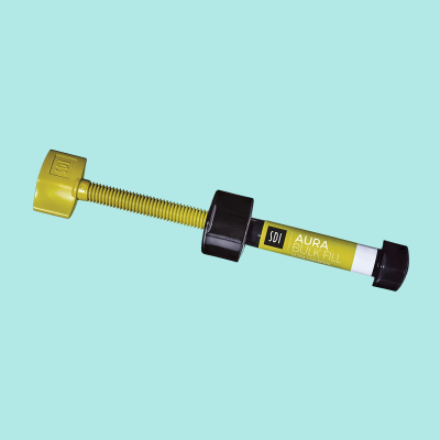
Effective Bulk Fill Composite
Bulk fill composites have seen a rapid rise in popularity over the years, this is your go to guide to know what makes a good bulk fill composite.
Kerr Restoratives
Designed to make life easier for dentists, the Artisan range of restorative solutions from Kerr Dental consists of award-winning products and formulas.
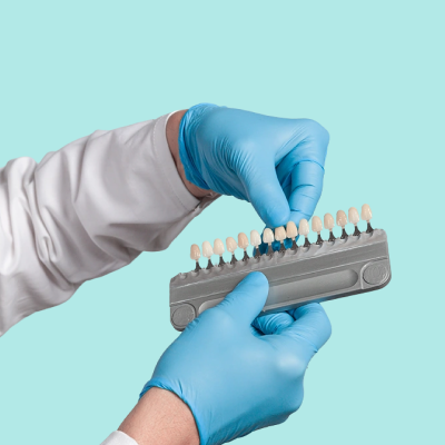
Universal Shade Composite
Minimise the number of shades needed in your practice with universal composite and discover the key benefits.

Hearing Loss in Dentistry
Most dentists would agree that their profession is a noisy one, but how many understand just how noisy it is or the health risks involved?

Dental Waste Disposal
Read Kent's guide to disposing dental waste safely and legally. Ensure your practice is fully compliant and optimising its waste management operation.
Guide to Endodontic Files
Find the perfect endodontic file for your needs with our comprehensive buying guide. From materials to sizes, we'll cover everything you need to know. Shop now.
Dental Extraction Products
Should you let patients take their extracted teeth home? Find out the benefits and risks with Kent Express' helpful guide. Shop now for dental extraction products.
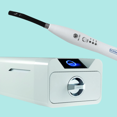
Testing Your Curing Light
See how radiometers and new age curing devices can eliminate doubt and help guarantee fully cured fillings every time.
Why Consider Dental Glass Hybrid
Find out more about the advantages of using dental glass hybrids as a restorative over composites and amalgam.
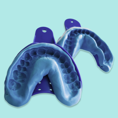
Best Dental Impression Material
Find out what the best dental impression material is for your practice with Kent Express' helpful guide. Shop a wide variety of addition silicone, condensation silicone, alginates and polyether in stock for next working day delivery
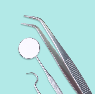
Guide to Dental Instruments
Find the right dental instruments for your practice with Kent Express' comprehensive buying guide. Get tips, recommendations, and top-quality products. Shop now.
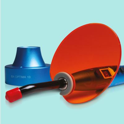
Dental LED Curing Light
Few pieces of equipment are used as frequently in the surgery as the dental curing light they comewith different features and clinical capabilities.
How to Make Cementation Simpler
Find out how 3M RelyX Universal can simplify and standardise cementation procedure.
Advantages Glass Ionomer Cement
Learn about the advantages of Glass Ionomer Cement (GIC) on Kent Express blog. Discover its uses, benefits, and how it compares to other dental materials.
Guide to Dental Composites
Looking for the best dental composites for your practice? Check out Kent Express' comprehensive buying guide, with tips and recommendations for every need.